M, N, O, P
Structure and Function of Neurons
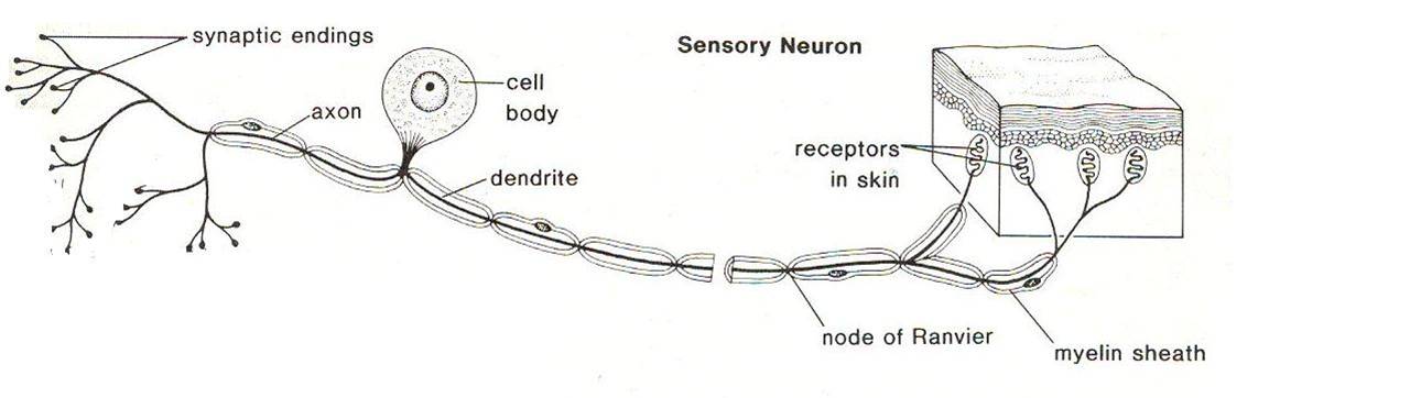
Sensory Neuron

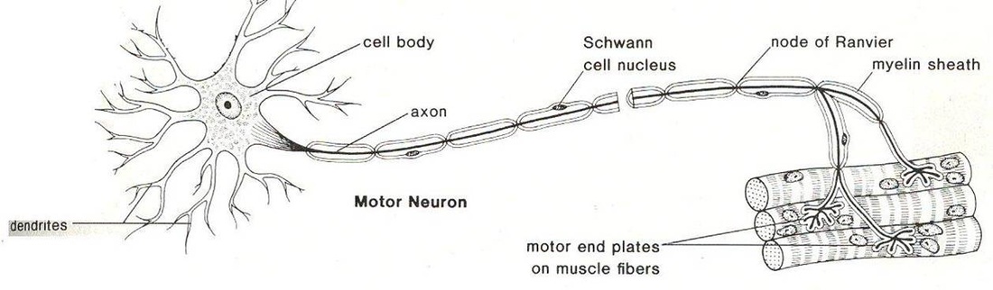
Motor Neuron
Interneuron

Sensory Neuron- The sensory neuron is located near the skin or organs, where ever there is stimulous. The sensory neurons are an afferent neuron meaning that they move away from a central organ or point. They relay the message from receptors to the brain or spinal cord.
Motor Neuron- The motor neuron is efferent, meaning it is relaying the message away from the brain and towards the central point or organ. They are located in the brain and spinal cord and they tell the central point or organ to react.
Interneurons- these are the neurons that connect the sensory neurons to the motor neurons
Parts of the Neuron
1. Dendrites -Conduct a nerve impulse (message) towards a cell body.
-Many dendrites enter a cell body.
2. Cell Body -Contains the nucleus and cell organelles needed to keep the cell alive.
-Relays impulse from Dendrite to Axon.
3. Axons -Conduct a nerve impulse away from the cell body.
4. Myelin Sheath -Protective coating of Schwann Cells around larger Axons
5. Nodes of Ranvie -Interrupted areas on the Myelin Sheath
-Speeds up transmission of impulse.
6. Synaptic Terminal
-Junction through which neurons signal to each other and to non-neuronal cells such as those in muscles.
Reflex Arc

The Reflex arc involves your bodies reflexes to events without sending the signal to your brain first. The reason the reflex arc doesnt include your brain is because it would take too long for you to react to emergencies. If you put your hand on a hot stove, you immediately remove it, without even thinking about it. If you did think about it, you may burn your skin and do some serious damage. Instead of going up to your brain, the sensory neurons detect the stimulus and send the signal up your interneurons, to the motor neurons located in your spinal cord. The motor neuron takes the message away from the central nervous system (located in your spinal cord) and towards the effector (site of stimulus). Then the muscle receives the message
and contracts. Senses like your eyes and ears let your brain know what happened after the fact.
Division ofNervous System

- Central- Brain, Spinal Cord
- Peripheral- Somatic, Autonomic
- Brain and Spinal cord- Neurons and supportive tissue, spinal column
- CNS- Sensory nerves (skin, muscle, some internal organs), Motor nerves
- PNS- Somatic (voluntary actions), Autonomic (Non voluntary)
- Sympathetic- Fight or Flight
- Parasympatheic- Rest and Digest
The Brain
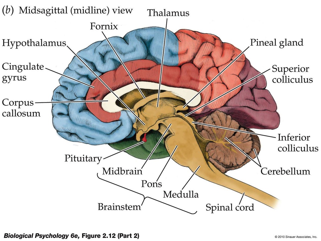
- BRAIN STEM: The part of the brain that connects to the spinal cord. The brain stem controls functions basic to the survival of all animals, such as heart rate, breathing, digesting foods, and sleeping. It is the lowest, most primitive area of the human brain.
- CEREBELLUM: Two peach-size mounds of folded tissue located at the top of the brain stem, the cerebellum is the guru of skilled, coordinated movement (e.g., returning a tennis serve or throwing a slider down and in) and is involved in some learning pathways.
- CEREBRUM: This is the largest brain structure in humans and accounts for about two-thirds of the brain’s mass. It is divided into two sides — the left and right hemispheres—that are separated by a deep groove down the center from the back of the brain to the forehead. These two halves are connected by long neuron branches called the corpuscallosum which is relatively larger in women’s brains than in men’s. The cerebrum is positioned over and around most other brain structures, and its four lobes are specialized by function but are richly connected. The outer 3 millimeters of “gray matter” is thecerebral cortex which consists of closely packed neurons that control most of our body functions, including the mysterious state of consciousness, the senses, the body’s motor skills, reasoning and language.
- THALAMUS: Located at the top of the brain stem, the thalamus acts as a two-way relay station, sorting, processing, and directing signals from the spinal cord and mid-brain structures up to the cerebrum, and, conversely, from the cerebrum down the spinal cord to the nervous system.
- HYPOTHALAMUS: Located at the base of the brain where signals from the brain and the body’s hormonal system interact, the hypothalamus maintains the body’s status quo. It monitors numerous bodily functions such as blood pressure and body temperature, as well as controlling body weight and appetite.
- Medulla Oblongata -Brain Stem (bottom of the brain)
-Pathway between brain and spinal cord
-Controls: Vomiting, coughing, sneezing,
hiccoughing, swallowing.
-Controls: Heartbeat rate, breathing, and
blood pressure.
Urinary System Functions
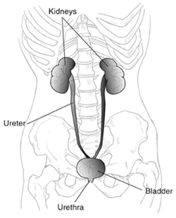
Kidney – Produce urine
Ureters – Transport urine to
the bladder
Bladder – Storage of urine
Urethra - Elimination of urine
Ureters – Transport urine to
the bladder
Bladder – Storage of urine
Urethra - Elimination of urine
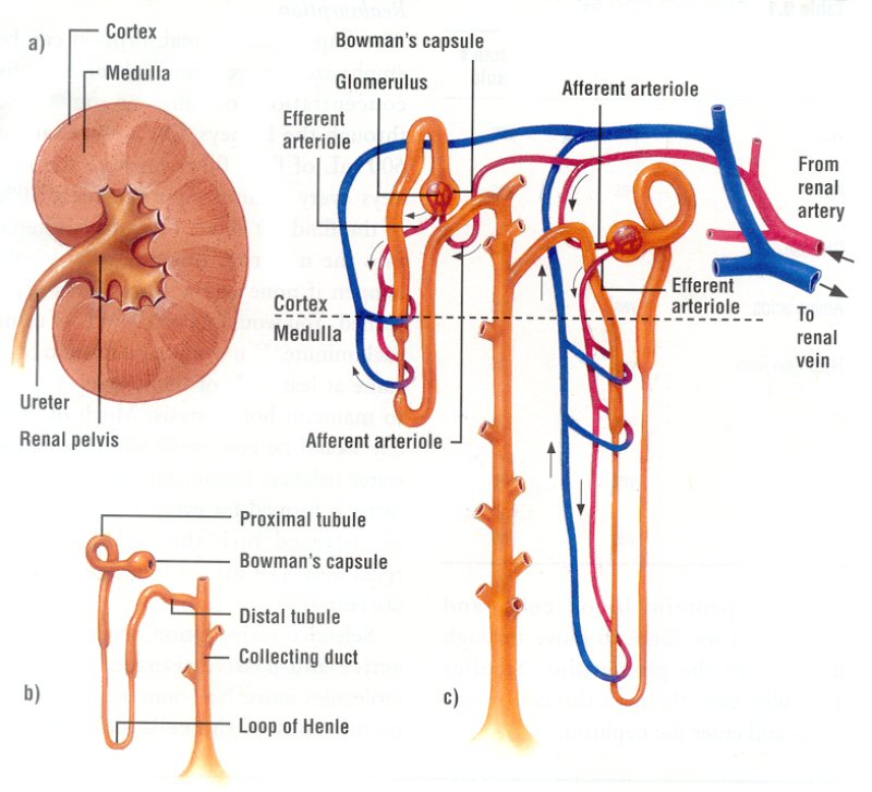
Production of Urine
When blood enters the glomerulus, pressure filtration happens. Water, nutrients and wastes are brought into the bowmans capsule. Large molecules dont enter the bowmans capsule, but stay in the blood system. If the pressure of the blood is too low, than filtration will not occur. To prevent this, a hormone called rennin is released inside the glomerulus, it constricts the glomerulus so the pressure is at a suitable level. At the Proximal Convultaed Tubules, more filtration happens. Required nutrients (glucose, amino acids, vitamins, minerals, salt molecules and some water) are reabsorbed into the peritubular capillary network. This process requires ATP to happen. Water must be reabsorbed into the blood since the body needs it to survive, so this is where the loop of henle comes in. The cells in the loop of henle pump out sodium to make it hypertonic. Once water is absorbed into the blood stream, aldosterone helps reabsorb the sodium). Water is reabsorbed making urine more concentrated inside the tract. Tubular secretion happens at the Distal Convulated tubules. histimine, penicillin, Hydrogen ions, and ammonia are secreted out of the blood into the tubules which helps maintain homeostacis. This helps maintain the pH of blood. Finally, at the collecting duct, water is reabsorbed into the blood. This makes the urine concentrated and it is ready to be excreted.
Male Reproduction

-Urethra -Conducts sperm out
of the body
-Ductus Vas Deferens- Conducts
and stores sperm
-Penis -Serves as an organ of copulation-Prostate Gland- A single dough-nut shaped gland that surrounds the upper portion of the urethra just below the bladder.
-Epididymus -Stores sperm as they mature
-Seminal Vesicle -Contributes to seminal fluid
-Cowper’s Gland -Pea-sized organs that lie posterior to the prostate on either side of the urethra.
-Contributes to seminal fluid
Makeup of Sperm

Female reproductive parts
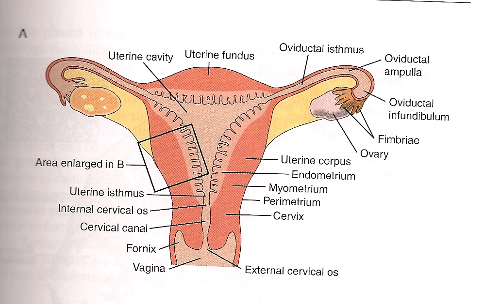
Ovaries - produce eggs and sex hormones
Uterus - houses fetus
Fallopian tube - conducts egg towards the uterus
Fimbriae - little finger things projecting from the Fallopian tubes which brush over the ovaries and help sweep the egg into the tube
Cervix - bottom of the uterus, leads to vagina. It dilates at birth to allow the baby through
Vagina - receives the penis and serves as a birth canal
